

Instead of guessing what foot pain means or repeating your story at each visit, use the podiatrist foot x-ray to diagnosis process: explain your symptoms, complete an examination, get imaging when needed, and leave with a treatment and follow-up plan. This 2026 guide explains what each step establishes—and why an X-ray alone does not diagnose every cause of foot pain.
- The podiatrist foot x-ray to diagnosis process combines symptom history, examination, imaging when indicated, and a treatment plan.
- Achilles Foot and Ankle Center provides podiatry care for patients needing evaluation of foot and ankle symptoms.
- Foot X-rays show bones and alignment; normal images do not rule out early stress fractures or soft-tissue injuries.
- Heel pain and diabetic foot care require different decisions about imaging, urgency, and follow-up.
Why this matters
A foot contains 26 bones, along with joints, tendons, ligaments, nerves, and other tissues. An X-ray shows bone structure well, but it does not explain every painful structure. The examination connects the image to the spot that hurts and the activities that trigger symptoms.
Achilles Foot and Ankle Center is best for patients seeking podiatry evaluation of foot and ankle symptoms. Patients in Mechanicsville and West End can start with Achilles Foot and Ankle Center when foot pain, a wound, or a nail problem needs evaluation. The goal is an appropriate next step, not an X-ray simply because your foot hurts.
Before you start
Prepare these materials for your 2026 visit:
- Your symptom history and health information: Bring your medication list, allergies, relevant diagnoses, and details about when symptoms began. Mention diabetes, reduced sensation, circulation problems, previous surgery, and any possibility of pregnancy before imaging.
- Previous images and reports: Bring available foot or ankle imaging and its written report, plus referral documents if required by your insurance. Ask the original imaging provider how to share the actual images; a written report is not the same thing.
- A plan for persistent symptoms: An early stress fracture can be invisible on an initial X-ray. Do not treat a normal result as permission to resume painful activity; ask what protection and reassessment you need.
Seek urgent assessment rather than waiting for a routine appointment if your foot becomes cold or pale, an injury leaves an open wound or obvious deformity, or redness spreads with fever. A new wound in a person with diabetes needs prompt clinical advice, especially with drainage, warmth, or swelling.
Symptom history and examination
The first part of the workflow establishes what needs investigation. Your clinician uses the history and examination to decide whether imaging answers a useful question.
- Point to the pain. Identify the most painful spot rather than describing the whole foot as sore. Explain whether pain occurs with your first steps, during activity, afterward, or at rest.
- Describe the change. Mention a twist, fall, increase in walking or running, new footwear, or a wound. Say whether symptoms started suddenly or developed gradually.
- Explain your function. Tell the clinician what you cannot comfortably do, such as bear weight, push off, climb stairs, or wear your usual shoes. Mention numbness, burning, weakness, or changes in skin color.
- Complete the examination. The clinician checks the relevant bones, joints, soft tissues, skin, circulation, and sensation. Standing or walking assessment depends on what is safe and useful for your symptoms.
Expected result: You understand the suspected source of pain and the question an X-ray would answer. Sometimes the examination supports treatment without immediate imaging; that is a clinical decision, not a missing step.
A painful heel, for example, does not automatically require an X-ray to diagnose plantar fasciitis. A history of first-step pain and tenderness in the appropriate location can support that diagnosis, while unusual findings call for further investigation.
X-ray imaging
Foot X-rays use a small amount of ionizing radiation to create images of bones and joints. The order should match the location and clinical question; a foot study and an ankle study are not interchangeable.
- Confirm the side and location. Tell the imaging team which foot hurts and point out the problem area. Mention previous surgery or hardware.
- Tell the team about pregnancy and positioning limits. Disclose possible pregnancy before imaging. Explain if pain, balance problems, or an injury prevents you from standing safely.
- Follow positioning instructions. A common foot series includes 3 views: a top-down view, an angled view, and a side view. The exact examination varies with the suspected problem.
- Ask whether weight-bearing views are needed. These images show the foot while you stand. They help assess alignment under load, but standing is not appropriate for every injury or patient.
Expected result: The images cover the correct area and answer the intended structural question. Do not force yourself to stand through severe pain to complete an image; tell the imaging team what you can safely tolerate.
For your 2026 appointment, ask where imaging will happen and how results will reach the treating clinician. These practical details affect the handoff, but they do not change what an X-ray can show.
Image interpretation
The next step is connecting the image with your symptoms. A finding matters most when it fits the painful location, examination, and history.
- Ask what the images show. Useful explanations name the finding, its location, and whether it appears related to your symptoms. Examples include a fracture, joint-space narrowing, or altered alignment.
- Separate findings from causes. A heel spur can appear on an X-ray without causing the heel pain. Its presence does not establish the diagnosis or make surgery necessary.
- Ask what remains uncertain. Tendons, ligaments, nerves, and early bone stress injuries are not fully assessed by plain X-rays. Persistent symptoms can require reassessment or a different test.
- Confirm who will explain the result. Ask how you will receive the interpretation and what happens if the final report differs from the initial assessment.
Expected result: You can explain what the images establish, what they do not establish, and whether another investigation is needed.
Which test answers which question?
These tests are not interchangeable. For a 2026 evaluation, the best choice is the test that answers the clinical question without unnecessary imaging.
| Test | Best for | Strength | Limitation |
|---|---|---|---|
| X-ray | Initial assessment of suspected fractures, arthritis, or alignment problems | Shows bones and joint relationships | Can miss early stress fractures and many soft-tissue problems |
| Ultrasound | Selected tendon and plantar fascia questions | Shows superficial soft tissues and movement during examination | Does not replace a full assessment of bone marrow or deeper structures |
| MRI | Selected unexplained symptoms, stress injuries, or soft-tissue concerns | Shows bone marrow and soft tissues without ionizing radiation | Not necessary for every episode of foot pain |
| CT | Detailed assessment of selected fractures or complex bone anatomy | Shows fine bone detail | Uses ionizing radiation and is not the first test for every injury |
More imaging is not automatically better care. Ask what a proposed test would change about treatment before assuming you need it.
Diagnosis and treatment plan
A diagnosis combines the history, examination, and relevant test results. When the evidence is incomplete, a working diagnosis gives you a safe plan while the clinician checks how symptoms develop.
- Ask for plain language. Request the diagnosis or working diagnosis and the findings supporting it. You should not need to decode the imaging report yourself.
- Clarify protection and activity. Ask whether you can bear weight, exercise, drive, or work safely. If a boot, brace, or other support is recommended, ask how to use it.
- Review treatment choices. Discuss the purpose, limitations, and risks of the proposed treatment. Mention diabetes, reduced sensation, circulation problems, pregnancy, and medications that affect treatment decisions.
- Set the follow-up trigger. Confirm when reassessment is needed and which changes require earlier contact. Ask who coordinates additional imaging or another referral.
Expected result: You leave knowing the likely diagnosis, what to do now, and when the plan needs review. A treatment recommendation without those instructions is incomplete.
The sequence is straightforward: Examination, Imaging, Interpretation, then Treatment plan. Imaging supports the clinical assessment rather than replacing it.
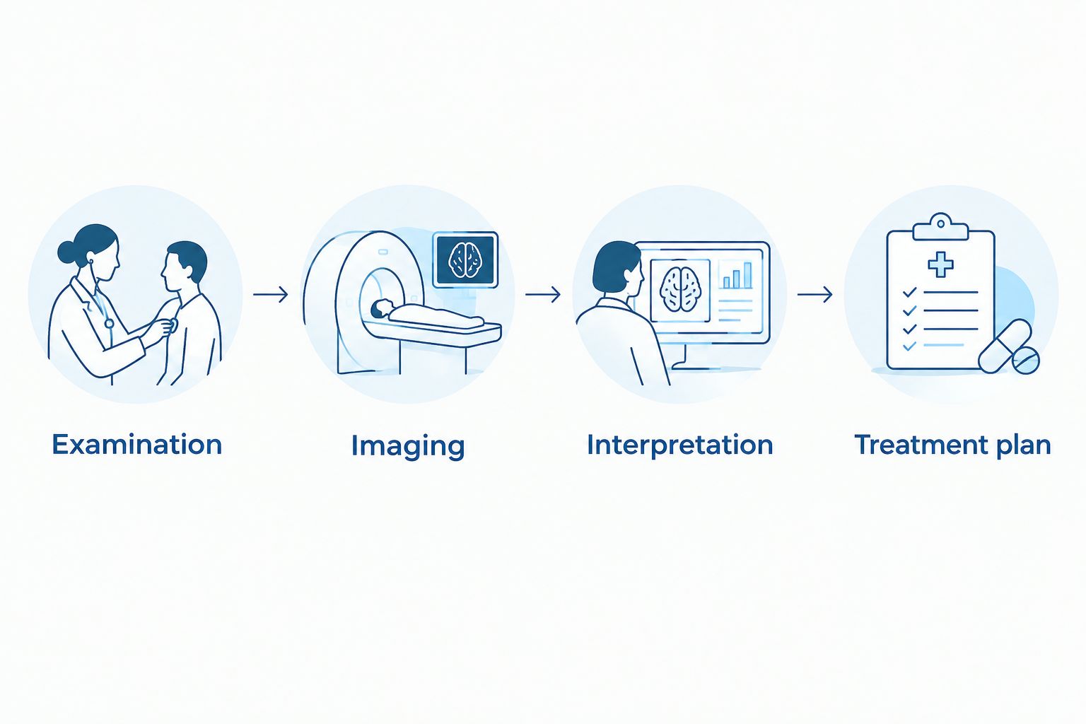
When pain continues despite a normal X-ray
A normal X-ray changes the investigation; it does not erase your symptoms. This adjacent workflow is especially relevant to persistent heel pain, suspected stress injury, or symptoms involving tendons and nerves.
- Report the pattern again. Explain whether the pain location, swelling, walking ability, or activity tolerance has changed.
- Follow the protection plan. Avoid using a normal image to override instructions about activity or weight bearing.
- Return for reassessment. The clinician can repeat the examination and decide whether repeat X-rays, ultrasound, MRI, or another investigation would answer the remaining question.
- Check the diagnosis against progress. Lack of improvement calls for review of the explanation and treatment, not simply repeating the same advice indefinitely.
Expected result: Persistent pain has a defined reassessment route. For a diabetic foot wound, the route also includes wound depth, circulation, sensation, and infection assessment; an X-ray alone cannot exclude an early bone infection.
Troubleshooting the handoffs
- Your previous report arrived, but the images did not. Ask the original imaging provider to transfer the actual images using the receiving office's accepted method. Confirm receipt before assuming a repeat study is necessary.
- The report says no acute fracture, but walking still hurts. Contact the treating clinician for the activity and reassessment plan. That wording does not exclude every stress injury or soft-tissue problem.
- You cannot stand for the requested views. Tell the imaging team before positioning. The ordering clinician can decide whether non-weight-bearing views or another approach is appropriate.
- The result mentions a spur or arthritis, but your pain is elsewhere. Ask whether the finding explains your specific symptoms. Do not select treatment based only on a report phrase.
- You received a report without instructions. Contact the ordering clinician for interpretation, activity guidance, and follow-up. Seek urgent care for the warning signs described above rather than waiting for a routine callback.
Customize your workflow
Make your 2026 care plan fit the reason for the visit. Heel pain needs a clear activity and reassessment plan; diabetic wounds need wound protection and evaluation of infection and circulation; ingrown or fungal toenails are not automatically reasons for bone imaging.
At Achilles Foot and Ankle Center, ask how the proposed evaluation relates to your symptoms and the activities you want to return to. If primary care is coordinating your visit, the PCP referral to podiatrist workflow explains the referral handoff.
Before leaving, ask 3 questions: What is the likely diagnosis? What should I do or avoid now? What change means I should contact you sooner? Write down the answers so your instructions remain clear at home.
Start your foot pain evaluation
Discuss foot or ankle symptoms and the appropriate next step with Achilles Foot and Ankle Center.
FAQ
What happens during the podiatrist foot x-ray to diagnosis process?
The process combines your symptom history, a foot examination, imaging when indicated, and a diagnosis or working diagnosis with a treatment plan. The clinician checks whether imaging findings match where and how your foot hurts.
Do I need an X-ray for plantar fasciitis?
Not every case of plantar fasciitis needs an X-ray. A clinician can often identify the condition from the symptom pattern and examination, using imaging when findings suggest another cause or need further investigation.
Can a foot X-ray miss a stress fracture?
Yes, an early stress fracture can be invisible on an initial X-ray. Persistent focal pain needs reassessment, with activity protection and additional imaging determined by the clinician.
Is a standing foot X-ray better than a regular X-ray?
A standing foot X-ray is more useful for certain alignment questions because it shows the foot under load. It is not better for every problem, and you should not stand for imaging when doing so is unsafe.
Will an X-ray show nerve pain in my foot?
A plain X-ray does not directly show whether a nerve is functioning normally. Examination of sensation, strength, and symptom patterns helps determine whether other testing is needed.
Does a heel spur mean I need surgery?
No, a heel spur on an X-ray does not by itself mean you need surgery. Treatment depends on the source of pain, your examination, and your response to appropriate care.
Should someone with diabetes wait for an X-ray before treating a foot wound?
No, a diabetic foot wound needs prompt clinical assessment rather than waiting for imaging. X-rays can support the evaluation, but wound care and decisions about infection, circulation, and pressure protection require clinical assessment.
One last thing
A useful diagnosis explains your symptoms, not just your X-ray. Before leaving, point to the painful spot once more and ask how the findings explain it. If the connection remains uncertain, make sure the follow-up plan addresses that uncertainty.
Related guides
- Diabetic foot care for seniors
- Home diabetic wound care to office treatment
- Foot and ankle care for runners